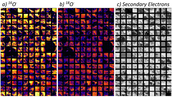
Micrometer-scale ion images of the cross-section of SaU 290

Micrometer-scale ion images of the cross-section of SaU 290 (CH3 chondrite), recorded by the NanoSIMS 50L at Academia Sinica. A ~10 pA Cs+ primary beam with a nominal spot size of 100 nm and rastered over 20 × 20 µm, to image the sample surface simultaneously in several ion species. A total of 160 (10 × 16 rows) individual images were recorded, and these were combined to create maps in a) 16O- and b) 18O-, as well as c) secondary electrons.